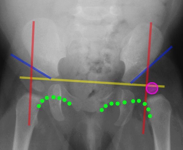
how do they x ray babies hips
The authors of the second paper also evaluated the amount of x-ray exposure during follow-up evaluation. DDH is more common in babies born in the breech position.
They determined that modern x-rays have very low dose exposure and the total of two pelvis x-rays was less than 001 mSv.

. Youll be asked to partly undress your baby and take off their diaper for the test. Appointments and Referrals. Hip X-rays are done with a child lying on a table.
In the direct projection or frontal obtained by focusing the x-ray tube perpendicular to the body plane - front or rear and axial transverse or horizontal plane fixing the elements of the joint from top to bottom - along the femur. Children use to grow and this means X-ray is done on baby bones and they will grow and growing tissue is altered by X-ray. A hip X-ray is a safe and painless test that uses a small amount of radiation to make images of the hip joints where the legs attach to the pelvis.
It is used for the assessment of unilateral hip pathology most commonly to diagnose a hip fracture or dislocation. Typically X-rays of both hips are taken for comparison even if only one hip is causing symptoms. Apparently baby X-rays are something known as a Pigg O Stat in which children between the ages of 12 months and 3 years old are put in for X-rays to keep them immobilized while also protecting them from exposure to harmful radiation.
First your baby will get medicine that makes them sleepy. During the examination an X-ray machine sends a beam of radiation through the pelvic bones and hip joints and an image is recorded on a computer or special film. During the test comfort your baby with a calm and soothing.
They should stay still for 23 seconds while each X-ray is taken so the. The maximum visual information is given by the x-ray of the hip joint in two projections. This image shows the soft tissues and the bones of the pelvis and.
A hip x-ray also known as a hip series or hip radiograph is a pelvis x-ray with an additional lateral view of the specified hip. How do they x-ray babies uk. Its used because babies and toddlers are incapable of following directions to hold still.
The doctor hears or feels a hip click when moving the infants thigh outward during a routine checkup. The American Academy of Pediatrics does not recommend routine ultrasounds for every infant. Your baby will be placed on a table on their back or side.
Hip X-rays are done with a child lying on a table. In these younger children it is possible to nip the problem early with night time bracing to help the hips recover if hip dysplasia returns. This is a basic article for medical students and other non-radiologists.
Youll be asked to partly undress your baby and take off their diaper for the test. If an image is blurred the X. Once the radiographer a person specially trained in taking X-ray images has positioned the part of your childs body to be examined and lined up the X-ray machine the X-ray examination takes less than a second to perform.
There are other less common factors that may. It might help to feed your baby just before the ultrasound to make your little one. You will go in the room with him he will need to be stripped from the waist down they will take x-rays of him flat on his back legs dead straight and together you wil be able to hold him in this position then an x-ray of his still on his back with his knees bent facing outwards and the soles of his feet put together he will be fine its not traumatic at all you.
But for babies with an abnormal physical exam or major risk factors for developmental dysplasia of the hip or DDH family history Breech position etc the AAP supports referral for. Also a picture can be taken with a lateral. X-ray examinations are usually quick and simple.
Usually both hips are scanned for comparison. This is about ten. Hip problems may not be present at birth.
Hip dysplasia is more common in first-born children because they are often more cramped in the uterus of first-time mothers. From the front anteroposterior view or AP from the side lateral view also known as the frog leg lateral view Typically X-rays of both hips are taken for comparison even if only one hip is causing symptoms. An X-ray technician will take pictures of the hip.
An X-ray technician will take pictures of the hip. It might help to feed your baby just before the ultrasound to make your little one more relaxed. They should stay still for 23 seconds while each X-ray is taken so the images are clear.
Your child may be required to hold his or her breath or remain still. Babies in the normal womb position have less pressure on their hips which is why they are far less likely to have DDH. The scan usually takes about 20 minutes.
Usually both hips are scanned.

Congenital Hip Dislocation Chd Happens When A Child Is Born With An Unstable Hip Read On To Learn Dislocacion De Cadera Huesos De La Cadera Batido De Papaya

Diapering And The Harness Cloth

Www Mdct Com Au On Instagram Anatomy Pathology Medicine Nursing Radiography Radiologictechnologist Radiology Radiologystudent Instagram Instagrammers

Pin On Congenital Adnormalities

Xray Image Child Swallowed Coins Medical Stock Photo 342401210 Shutterstock

Severe Hip Dysplasia In A Boxer The Red Arrows Are Pointing To The Over Growth Of Bone At The Femoral Neck Head The Red Arrow Shades Of Grey Hip Dysplasia

Pin On Adult Hip Dysplasia Awareness

How Parents Can Help Their Baby S Hip Dysplasia

What Causes Hip Dysplasia International Hip Dysplasia Institute


